Confocal fluorescence microscopy of enamel cells from developing rat tooth
Histology
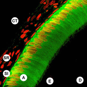
The principal enamel cell is termed 'ameloblast'. The ameloblast layer (A) faces developing enamel (E; not visible in this image) and is backed by accessory epithelial cells termed 'stratum intermedium' (SI) and 'stellate reticulum' (SR). Surrounding vascularised loose connective tissue (CT) was removed during microdissection. The dentine (D) core of the tooth underlies the enamel layer and likewise is not visible in this image.
Dual colour confocal laser scanning microscopy
A cryosection of rat enamel epithelium (secretion phase) was labelled with antibodies to a calcium-binding protein (calbindin-28kDa, digitally coded as green) and with a DNA stain (propidium iodide, coded red). Ameloblasts contain high levels of calbindin in the cytosol (green) and lesser amounts in the nuclei (red/yellow). The accessory cells lack calbindin so only their red nuclei are seen. No calbindin or DNA was detected in the developing enamel and dentine (black).
Credit
Image by Chris Turnbull and Mike Hubbard (see: Sayer, R.J., et al (2000) [Medline reference]
Immunohistochemistry of developing rat tooth

Immunohistochemistry
Light microscopy of a paraffin section of developing rat molar labelled with antibodies to ERp29 (an endoplasmic reticulum resident) and counterstained with haematoxylin. The principal enamel cells (ameloblasts) contain high levels of ERp29 (dark brown) compared with the other cell types present. ERp29 is not detected in the developing hard tissues (enamel and dentine matrix).
Credit
Image by Steve Shnyder and Mike Hubbard. (see Shnyder SD and Hubbard MJ 2002 [Medline])
|